
Why electrons
A light microscope can only resolve two points as separate if they are further apart than about half the wavelength of light (Abbe):
d ≈ λ / (2 · NA) → with λ ≈ 400–550 nm and NA ≈ 1.4: d ≈ 150–200 nm
Viruses, ultrafine dust and nanoplastics are often considerably smaller. A virus measures about 100 nanometres.
Electrons have a wavelength that decreases with the accelerating voltage U:
λ ≈ 1.23 nm / √(U / V) → at 5 kV: λ ≈ 17 pm
That is around ten thousand times less than for visible light, so the wavelength practically no longer limits the resolution. What matters is how finely the electron beam can be focused.
How an image is formed
A scanning electron microscope does not take a photograph. At the top, a source emits electrons. Lenses focus them into a very fine beam, and a deflection system moves it across the sample: point by point, line by line, like the beam in an old television tube.
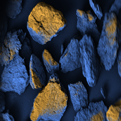
At each point, a detector counts how many electrons come back. Many means bright, few means dark. Heavy materials such as metals send back more electrons than light ones such as plastics – so brightness also reveals what a particle is made of.
Image quality is a matter of counting electrons: few electrons per point give a noisy image, many give a clear one – and that takes time. This is exactly where our AI comes in.
Why a vacuum – and our window
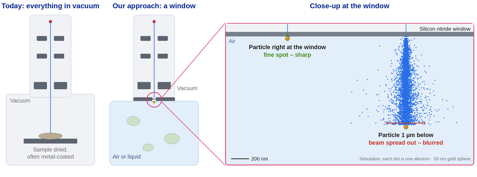
Electrons do not get far in air: within a fraction of a millimetre they collide with gas molecules and are deflected. That is why the sample in conventional electron microscopes sits in a vacuum. This comes at a price: the sample is dried and often coated with metal – anything living, moist or moving is lost, and a laboratory is a must.
Our approach is a window: a membrane of silicon nitride, much thinner than a hair, that separates the vacuum from the sample. The beam passes through it, and the sample stays in air or liquid – with no vacuum at the sample and no sample preparation.
The catch: right at the window the beam stays fine and the image sharp. The further a particle is from the window, the more the beam spreads out. Our physical model predicts by how much – electron by electron. This gives us the design rules for the instrument, and part of the blur can be recovered computationally.
Layout of the beam path
We develop our electron optics ourselves. The beam path follows the principle of the scanning electron microscope, from the source to the image:
- Thermionic cathode A thermal electron source.
- Wehnelt cylinder Shapes the emerging electron cloud and focuses it into a first crossover.
- Anode Accelerates the electrons.
- Electromagnetic lens Focuses the beam onto the sample. Today we work with an electromagnetic lens system; our long-term goal is electrostatic lenses, which can be built even more compactly.
- Deflection Moves the beam horizontally and vertically, line by line, across the sample – controlled by our own electronics.
- Detector and image formation The signal from each scan point builds up the image.
Design and physical model
Electron lenses can be calculated much like optical lenses. We determine the field distribution of a lens system with the finite element method (FEMM); the focal length follows from the field along the axis of rotation (after Reimer). We have cross-checked the design with several independent calculation methods.
Today we work with an electromagnetic lens system. Electrostatic lenses are our long-term goal: they can be built even more compactly.
On top of this comes a physical model of the entire instrument: a Monte Carlo calculation that follows the beam electron by electron through window, air and sample, validated against published measurements. It predicts what the instrument can see – and provides training images for which we know the correct answer.
Testing in high vacuum
Every version of the beam system is examined for its electrical properties in a vacuum chamber, first in a bell jar with a fluorescent screen on which the beam diameter can be observed. Simulation and measurement are compared until the model reliably predicts the design.
For this we operate high-vacuum test rigs with turbomolecular pumps and scanning electron microscopes in our laboratory in Künzelsau.
Physical AI
Almost anyone can use AI software today. The advantage lies with those who combine it with their own measuring hardware. At MeiLuft, the AI works on two levels:
- In the image. It improves noisy and blurred images and is designed to recognise each individual particle by shape and material. It is trained on images from our physical model for which we know the correct answer – the only way to measure whether it really finds more particles or merely paints prettier pictures.
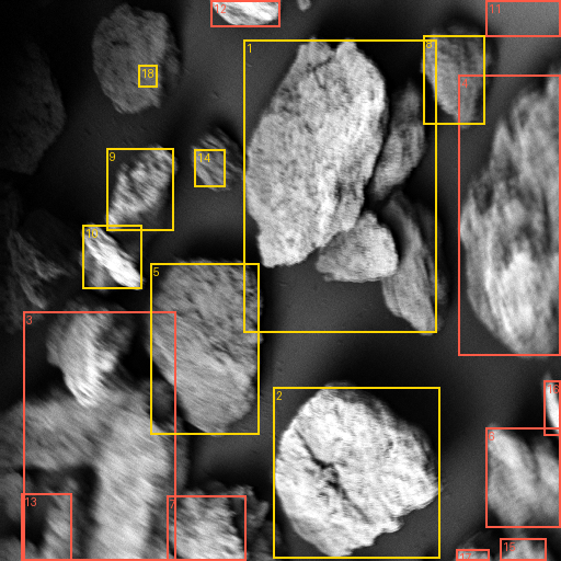
- At the instrument. It is designed to run the microscope itself – down to the level of the coils and the scan signal, with our own electronics and software. Our test microscope already scans with this control system and finds particles on its own, for now with classical image processing. The next step is a research agent that plans and carries out measurements.
AI agents on electron microscopes are a young field of research; the first studies rely on commercial instruments and their manufacturer interfaces. What sets us apart: our own hardware right down to the beam, a physical model of the instrument – and a microscope that measures in air.
First images
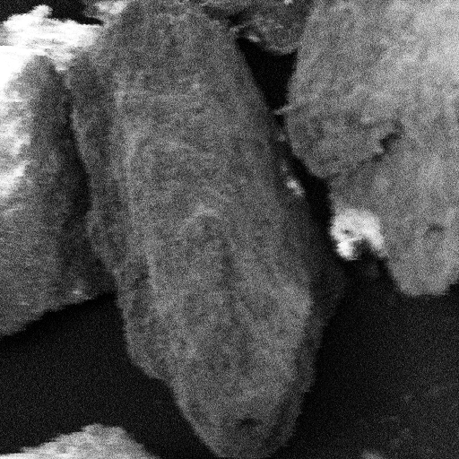
Recorded with our test microscope and our own instrument control. The instrument is not yet calibrated, which is why the images carry no scale bar.
Context for researchers
Electron microscopy outside high vacuum is not a new goal. Three designs have emerged:
- Closed wet capsule. The sample sits in a capsule behind a membrane, inside an otherwise conventional microscope (Thiberge et al. 2004).
- Inverted microscope with an open dish. The column looks up through a silicon nitride membrane into an open sample dish at atmospheric pressure (ASEM; Nishiyama et al. 2010). This was used to detect nanoparticles from about 30 nm directly in liquid (Luo et al. 2013).
- No specimen chamber at all. Electron microscopy directly in air (AirSEM; Nguyen, Holtz, Muller 2013).
In addition, there is liquid-cell transmission electron microscopy (review: de Jonge & Ross 2011) and environmental SEM (ESEM), which works with residual gas but not at full atmospheric pressure.
What MeiLuft does differently:
- Compact and in air – intended for use where the particles occur, not only in the laboratory.
- Our own hardware right down to the beam: our own electron optics, our own electronics and software for scanning and signal acquisition.
- A physical model as the foundation: it predicts what the instrument can see and provides training data with a known solution.
- Physical AI: AI that does not just analyse but operates the microscope. Agentic AI on electron microscopes has been a visible field of research since 2026 (for example Wall et al. 2026; Zhang et al. 2026) – so far on commercial instruments via manufacturer interfaces.
Open questions we are working on:
- How far may a particle be from the window and still remain sharp?
- How fast may a particle move and still be imaged?
- How much can a physics-informed AI recover from noisy, blurred images – and how can this be tested honestly when there is no clean reference image?
- S. Thiberge et al.: Scanning electron microscopy of cells and tissues under fully hydrated conditions. PNAS 101 (2004) 3346–3351. doi:10.1073/pnas.0400088101
- H. Nishiyama et al.: Atmospheric scanning electron microscope observes cells and tissues in open medium through silicon nitride film. J. Struct. Biol. 169 (2010) 438–449. doi:10.1016/j.jsb.2010.01.005
- N. de Jonge, F. M. Ross: Electron microscopy of specimens in liquid. Nat. Nanotechnol. 6 (2011) 695–704. doi:10.1038/nnano.2011.161
- P. Luo et al.: Visualization and characterization of engineered nanoparticles in complex environmental and food matrices using atmospheric scanning electron microscopy. J. Microsc. 250 (2013) 32–41. doi:10.1111/jmi.12014
- K. Nguyen, M. Holtz, D. Muller: AirSEM: Electron Microscopy in Air, without a Specimen Chamber. Microsc. Microanal. 19 (S2) (2013) 428–429. doi:10.1017/s1431927613004133
- M. K. Wall et al.: TEM Agent: enhancing transmission electron microscopy with modern AI tools. npj Comput. Mater. 12, 316 (2026). doi:10.1038/s41524-026-02103-z
- K. Zhang et al.: AutoRASOR: Autonomous Rapid Scanning Electron Microscope Operator. arXiv:2609.22411 (2026). arxiv.org/abs/2609.22411
Are you working on electron optics, sample delivery or image analysis and would like to collaborate with us? Write to us at durchatmen@meiluft.de.
Glossary
- Scanning electron microscope (SEM)
- Scans a sample point by point with a fine electron beam and assembles an image from it.
- Backscattered electrons
- Electrons reflected back by the sample. Heavy materials reflect more – this creates material contrast.
- Window (membrane)
- Electron-transparent membrane of silicon nitride that separates the vacuum from the sample.
- Nanometre
- One millionth of a millimetre. A virus measures about 100 nanometres, a hair around 50,000 to 100,000.
- Ultrafine dust
- Airborne particles below 100 nanometres.
- Nanoplastics
- Plastic particles below one micrometre.
- Physical AI
- AI that does not just analyse data but controls hardware.
- Physical model
- Also called a digital twin: a calculation that predicts what the instrument will see – electron by electron.
Frequently asked questions
Is this an electron microscope?
Yes – a compact scanning electron microscope in which the sample does not have to go into a vacuum.
Why not simply use a light microscope?
Light cannot resolve anything smaller than about 200 nanometres. Many of the particles that matter are smaller.
Doesn’t this already exist?
In part. Electron microscopes with a window exist in research, for example for samples in liquid. What is new is the combination: compact, in air, with our own control right down to the beam and an AI that improves images, recognises particles and operates the instrument itself.
When will the instrument be available?
We are building the prototype; how fast it goes depends on funding.
How small can it see?
We will show that with the prototype.